
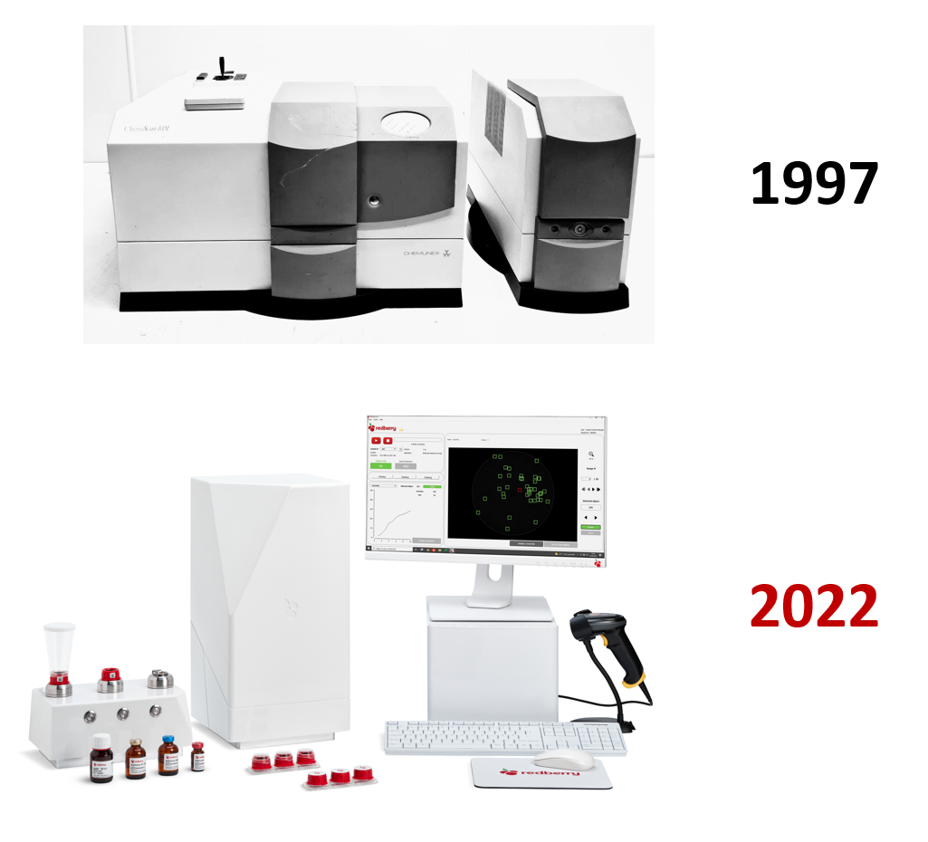
The Genesis of SPC at Chemunex
Solid Phase Cytometry (SPC) was developed in the late 1990s by the French company Chemunex to meet the growing demand for rapid and sensitive microbial detection. The concept of single-cell detection was not entirely new: the Direct Viable Count (DVC) method [1] had already shown this possibility in the 1970s, but its sensitivity (~10⁴ cells) was limited by microscopic observation of only a few fields (typically about ten per membrane). SPC overcame these limitations by combining membrane filtration, fluorescent viability staining, laser scanning, and digital image analysis to scan the entire filter surface.
SPC was conceived and developed through a series of innovations and patents led by Dr. Jean-Louis Drocourt, who played a central role in its industrial implementation. Notably, Chemunex was later acquired by AES, possibly as a consequence of financial difficulties linked to the high development costs of the ChemScan platform. This acquisition ensured the technology’s continuation and evolution in industrial settings.
Before its application to sterile pharmaceutical products, SPC was used in diverse environmental and industrial contexts. It enabled total viable counts in water [2,4,6], detection of fecal indicators such as E. coli [3,4] and Enterobacteriaceae [6], and enumeration of pathogens like Legionella [8] and Cryptosporidium [2]. It also detected airborne fungi, including Aspergillus fumigatus, in hospital and cleanroom settings [10].
These early studies [2–10] demonstrated SPC’s robustness in detecting low microbial loads across many matrices and laid the foundation for later pharmaceutical applications where speed and sensitivity are critical.
Unique and still unmatched performance
SPC quickly proved highly performant, combining high sensitivity with the ability to analyze individual microbial cells in situ. Using CFDA-based viability probes that label only intact, metabolically active cells [5], SPC achieves a detection limit as low as 1 CFU per sample, with results in 2–5 hours [2–11].
Relying on viability markers, SPC detects VBNC cells missed by culture, with imaging algorithms ensuring accurate discrimination; accordingly, studies consistently report higher counts.
Compared to other direct methods such as ATP bioluminescence [12] and flow cytometry [13], SPC offers much greater sensitivity and specificity. ATP-metry typically detects only 10³–10⁴ CFU/mL, making it useful for trend monitoring but unsuitable for low-level contamination without enrichment. Flow cytometry can resolve individual particles but often struggles to separate viable cells from debris or autofluorescence, with practical thresholds rarely below 10² CFU/mL. Unlike these methods, SPC delivers true single-cell viable detection—combining sensitivity, specificity, and speed at a level unmatched by alternative direct approaches.
Unique and still unmatched performance
SPC rapidly established itself as a method offering exceptional analytical performance. It combines high sensitivity with the ability to analyze individual microbial cells in situ. Using CFDA-based viability probes, which label only viable cells with intact membranes and active enzymatic metabolism, SPC achieves a limit of detection as low as 1 CFU per sample, with results available in 2 to 5 hours [7, 8].
The method does not rely on microbial growth, enabling detection of viable but non-culturable (VBNC) cells that would otherwise escape standard plate-based methods [1, 9]. Its imaging and signal-processing system ensures accurate identification by analyzing spot size, intensity, color ratio, and signal pattern. In side-by-side comparisons, SPC often yielded higher microbial counts than classical culture, highlighting its sensitivity and its ability to account for the full viable population [7].
Why SPC was not widely deployed despite its performance ?
Despite its strengths, several practical limitations initially prevented SPC from widespread routine use in industrial microbiology. The analytical workflow required multiple manual steps—filtration, staining, incubation, rinsing, drying, and loading of the membrane into the scanner—followed by microscopic confirmation of each fluorescent event with a motorized epifluorescence microscope. This final step introduced operator dependency, slowed the process, and created a bottleneck in high-throughput settings.
Open handling steps further increased the risk of contamination and false positives, a critical concern in aseptic environments. Samples with high background fluorescence or particulates often led to aborted scans or labor-intensive validation. As the FDA has noted in 2019 [14], these technical limitations—combined with high cost, large footprint, and limited throughput—confined SPC’s adoption to niche applications such as rapid sterility testing for short shelf-life products.
Filterability was never the real limitation…
Despite common belief, filterability has never been a real barrier for SPC. Many so-called “non-filterable” matrices were successfully processed using suitable pretreatment protocols or membrane configurations. SPC has been applied to a wide range of challenging samples, including milk [9], pharmaceutical oils, bronchoalveolar lavage (BAL), sputum, and surface waters [6]. It enabled detection of Mycobacterium paratuberculosis in spiked milk [9], Naegleria fowleri in natural waters [6], and Aspergillus fumigatus in respiratory fluids and ambient air [10]. Filtration was achieved either directly or after steps such as polymer dissolution or magnetic capture, and even in turbid matrices SPC provided sensitive and specific detection.
More recently, its applicability was confirmed in advanced therapy products, where SPC enabled rapid and reliable microbial testing [17].
These results underscore the method’s versatility beyond clear aqueous solutions.


Making SPC Ready for Routine Use: Redberry’s Contribution
Recent developments have addressed the limitations that long hindered the broad adoption of SPC.
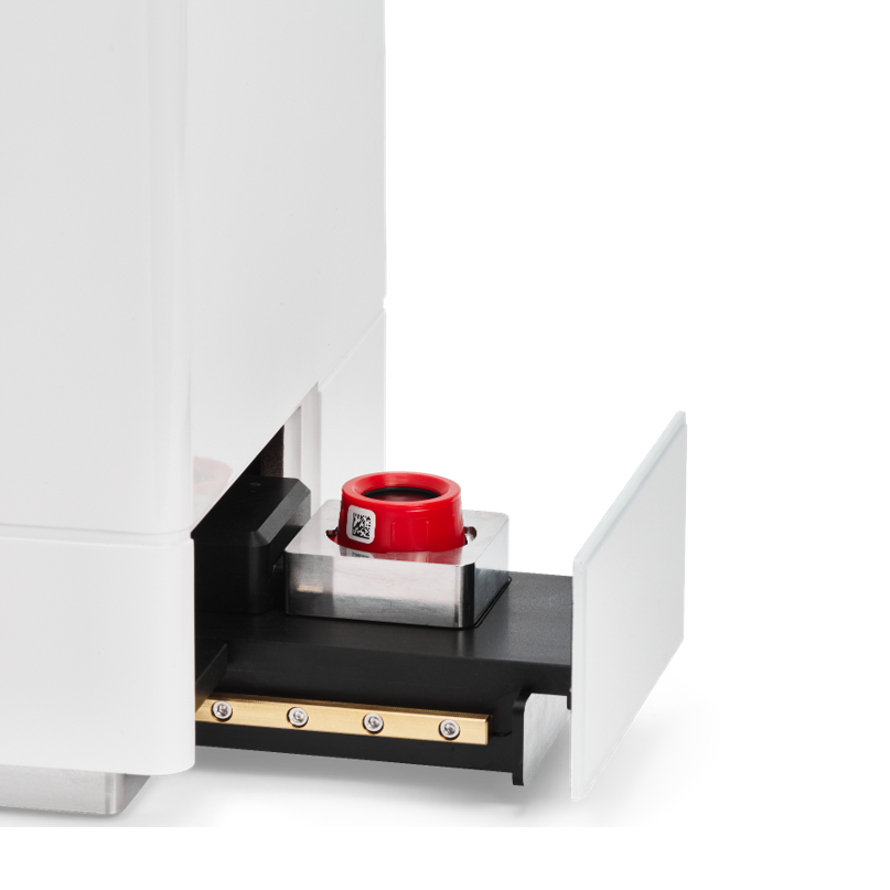
In 2021, Redberry introduced the Red One™ platform, a fully automated SPC system that integrates filtration, staining, detection, and result interpretation within an aseptic workflow [15–17]. A key innovation is the use of fluorescent staining kinetics, which enables automated discrimination of viable microorganisms and eliminates the need for manual microscopic confirmation.
The platform offers a rapid screening mode that delivers total viable cell counts in about 10 minutes across sample volumes from a few microliters to hundreds of milliliters, with sensitivity down to only a few viable cells. Beyond this first-level test, it performs quantitative bioburden assessments in roughly 4 hours, matching compendial accuracy without subculture or enrichment. For sterility testing, it provides results within 4 days while still allowing microbial identification. This workflow has been validated under both the European and U.S. Pharmacopeias, offering a GMP-compliant alternative to the traditional 14-day method and supporting faster product release.
The system also applies to complex biological matrices, including cell and gene therapy products, using selective lysis protocols to remove mammalian cell interference without compromising microbial detection. Compact, calibration-free, and fully aligned with data integrity requirements (21 CFR Part 11), Red One™ establishes SPC as a routine, validated, and regulatory-accepted technology.
By overcoming the last barriers to adoption, Redberry has enabled Solid Phase Cytometry to fulfill the vision set more than two decades ago: a method combining speed, sensitivity, and compliance for modern microbiological control.
Making SPC Ready for Routine Use: Redberry’s Contribution
Recent developments have addressed the limitations that long hindered the broad adoption of SPC.
In 2021, Redberry introduced the Red One™ platform, a fully automated SPC system that integrates filtration, staining, detection, and result interpretation within an aseptic workflow [15–17]. A key innovation is the use of fluorescent staining kinetics, which enables automated discrimination of viable microorganisms and eliminates the need for manual microscopic confirmation.
The platform offers a rapid screening mode that delivers total viable cell counts in about 10 minutes across sample volumes from a few microliters to hundreds of milliliters, with sensitivity down to only a few viable cells. Beyond this first-level test, it performs quantitative bioburden assessments in roughly 4 hours, matching compendial accuracy without subculture or enrichment. For sterility testing, it provides results within 4 days while still allowing microbial identification. This workflow has been validated under both the European and U.S. Pharmacopeias, offering a GMP-compliant alternative to the traditional 14-day method and supporting faster product release.
The system also applies to complex biological matrices, including cell and gene therapy products, using selective lysis protocols to remove mammalian cell interference without compromising microbial detection. Compact, calibration-free, and fully aligned with data integrity requirements (21 CFR Part 11), Red One™ establishes SPC as a routine, validated, and regulatory-accepted technology.
By overcoming the last barriers to adoption, Redberry has enabled Solid Phase Cytometry to fulfill the vision set more than two decades ago: a method combining speed, sensitivity, and compliance for modern microbiological control.
